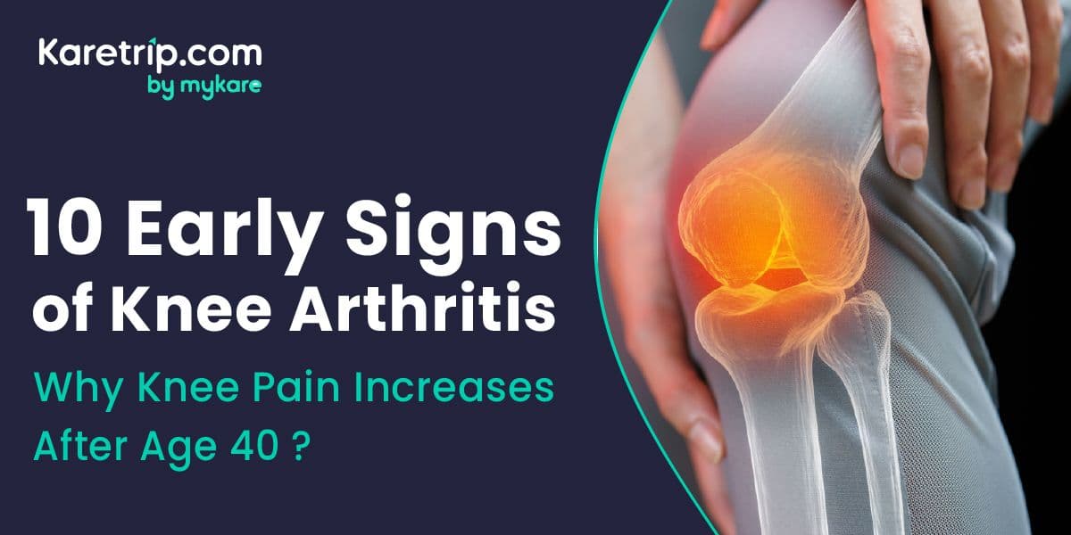

You used to run for the bus without thinking. Now, you hesitate before stepping off a curb. For many people, turning 40 brings an unwelcome birthday gift: a subtle, nagging ache in the knees. It’s easy to dismiss it as "just getting older" or "sleeping funny." But more often than not, this is the body’s check-engine light flickering on.
It is the start of Osteoarthritis (OA), the "wear and tear" disease.
Think of the cartilage in your knee like the tread on a car tyre. After decades of walking, running, and carrying weight, that tread wears thin. When the cushioning is gone, the bones start to complain.
Recognizing the early symptoms of knee arthritis can save you from surgery down the road. If you catch it now, you can preserve the joint. If you ignore it, the damage becomes permanent.
Here are the 10 specific signs that your knees are asking for help.
Why Knee Pain Specifically Increases After Age 40: The Biology
Before diving into the symptoms, it is worth understanding why 40 is the inflection point rather than 35 or 55. Three distinct biological changes converge at this age and their combined effect on the knee is significant.
1. Chondrocyte Decline
Cartilage is not bone it has no blood supply, no nerves, and no direct lymphatic drainage. This makes it exceptional at load-bearing but extremely limited in its ability to self-repair. The cells responsible for producing and maintaining cartilage are called chondrocytes, and they begin declining in both number and repair capacity from around age 40 onward.
Aging chondrocytes produce progressively less Type II collagen and aggrecan the two structural proteins that give cartilage its shock-absorbing, compressive properties. They also become increasingly vulnerable to oxidative stress, meaning that physical loading which a younger person's cartilage would absorb and recover from begins to leave permanent micro-damage after 40. The result: the cartilage is still there, but it is slowly becoming less capable of renewing itself between stresses.
2. Synovial Fluid Changes
The knee joint is enclosed in a capsule lined with the synovial membrane, which produces synovial fluid the joint's natural lubricant and nutrient source for cartilage. After age 40, synovial fluid gradually becomes less viscous and lower in hyaluronic acid concentration. This reduces the fluid's ability to evenly distribute load across the joint surface and keep the cartilage adequately lubricated during movement.
Less lubrication means more friction. More friction means faster cartilage wear. And less nutrient delivery via synovial fluid means slower cartilage recovery between loading episodes.
3. Sarcopenia (Muscle Mass Loss)
Muscle mass loss begins quietly from around age 30–35 and accelerates meaningfully after 40 at approximately 1–2% per year without active resistance training. For the knee, the quadriceps group (particularly the vastus medialis oblique, or VMO) is the primary muscular shock absorber. A healthy quadriceps can absorb a significant proportion of ground reaction force before it reaches the joint surface.
As quadriceps strength declines, more of that force travels directly to the cartilage, menisci, and bone. The joint was not designed to absorb this load unassisted and it complains accordingly.
The 10 Early Signs of Knee Arthritis
1. The "Morning Rust" (Stiffness)
This is the classic hallmark of early-stage knee arthritis. When you first wake up or sit at your desk for an hour, your knees feel stiff, like a rusty hinge that needs oil.
The tell-tale sign: You have to shuffle for the first few steps before you can walk normally. Unlike muscle soreness, this stiffness usually loosens up after 30 minutes of movement.
2. The "Staircase Struggle."
Walking on flat ground might be fine, but stairs are a nightmare. This is often the very first of the knee osteoarthritis symptoms people notice.
Why it happens: Climbing stairs puts pressure of nearly 4x your body weight on the knee. If your cartilage is thinning, your knee simply can't handle that load anymore.
3.The "Crunchy" Sound (Crepitus)
Do your knees pop, crack, or grind when you squat? Doctors call this Crepitus. It is the sound of rough surfaces rubbing together. Healthy cartilage is slick like ice; arthritic cartilage is rough like sandpaper. If that noise comes with pain, it is a definitive sign of arthritis in the knees.
4. The Weather Forecast Knee
It sounds like an old wives' tale, but it is real. Many patients notice their knee joint arthritis symptoms flaring up when it rains or the temperature drops.
The Science: A drop in barometric pressure causes tissues in the joint to expand slightly. In an inflamed knee, this expansion increases pressure and pain.
5. Swelling Without Injury
Did your knee puff up even though you didn't bump it? This is "Water on the Knee." When the cartilage is irritated, the joint produces excess fluid to try and protect itself. If you notice one knee looking slightly larger or "puffy" compared to the other, it’s a red flag.


6. Instability (The "Giving Way" Feeling)
You are walking along, and suddenly, you feel like your knee is going to buckle or fold underneath you. This "giving way" sensation happens because the muscles around the joint (quadriceps) weaken as pain stops you from using them fully. It creates a dangerous cycle where you fear walking, leading to even weaker muscles.
7. Loss of Full Range of Motion
Try this test: Lie on your back and try to pull your heel all the way to your buttocks. If you can't bend it as far as you used to, or if you can't straighten your leg completely flat on the bed, you are losing "Range of Motion." Bone spurs (tiny jagged growths) might be physically blocking the joint from moving.
8. Pain That Disrupts Sleep
In the early stages, pain usually vanishes when you rest. However, as the condition progresses, you might find a throbbing ache keeps you awake at night. This is a sign that the inflammation is active even when you aren't moving.
9. Tenderness to Touch
Press your finger along the joint line (the gap between the upper and lower leg bones). In a healthy knee, this shouldn't hurt. If you have knee arthritis symptoms, this area will feel tender or sore, confirming that the inflammation is inside the joint capsule, not just in the muscle.
10. The Start of a Deformity
This is a later sign, but it starts slowly. As the cartilage wears down unevenly (usually on the inside of the knee), you might notice your legs starting to look "bow-legged" or "knock-kneed." Your alignment is physically shifting.
Non-Surgical Knee Arthritis Treatment Options in India
The most important message after a Grade 1–3 OA diagnosis: there is a significant treatment spectrum between "take paracetamol and hope" and "book your knee replacement." Here is what that spectrum actually looks like — and what each option costs in India versus the West.
Physiotherapy — The Most Evidence-Based Non-Surgical Intervention
Structured physiotherapy is the highest-evidence, lowest-risk intervention for Grade 1–3 knee OA. A physiotherapist-designed program specifically for OA focuses on:
-
Quadriceps strengthening: Particularly the VMO (the inner quad), which provides medial knee stability. Straight-leg raises, terminal knee extensions, and wall squats at controlled depth are standard exercises.
-
Hamstring and calf flexibility: Tight posterior chain muscles increase flexion resistance at the knee and worsen posterior capsule stress.
-
Proprioception training: Balance boards, single-leg stands, and perturbation training restore the neuromuscular stability disrupted by cartilage damage.
-
Gait retraining: A physiotherapist can identify and correct gait abnormalities that concentrate load on the damaged compartment.
NSAIDs and Topical Anti-Inflammatory Medications
Oral NSAIDs (naproxen, ibuprofen, diclofenac, celecoxib) reduce both pain and inflammatory mediator production within the joint. For Grade 2 OA, regular low-dose NSAIDs (under medical supervision with gastric protection) can significantly improve functional capacity.
Topical NSAIDs particularly diclofenac 1% gel applied directly to the knee have shown comparable local efficacy to oral NSAIDs for knee OA with significantly lower systemic side effects. Topical capsaicin cream (from chili pepper) provides additional analgesic effect through substance P depletion.
Corticosteroid Injections
A direct injection of corticosteroid (typically triamcinolone or methylprednisolone) into the knee joint provides powerful, rapid anti-inflammatory relief. The onset of action is typically within 48–72 hours, and the benefit lasts approximately 6–12 weeks.
Corticosteroid injections are most appropriate for Grade 2–3 OA with significant inflammatory flares. They are not a long-term solution repeated injections at short intervals may accelerate cartilage degradation but for managing a severe flare, preparing for a specific event, or buying time while physiotherapy takes effect, they are highly effective.
Hyaluronic Acid Injections (Viscosupplementation)
Hyaluronic acid is the primary component of healthy synovial fluid. Its concentration and molecular weight decline in OA-affected joints, reducing lubrication. Intra-articular hyaluronic acid injections given as a course of 3–5 weekly injections aim to restore joint lubrication and reduce friction-related pain.
The evidence is stronger for some patient groups than others Grade 2–3 OA patients who have not responded fully to NSAIDs and physiotherapy show the most consistent benefit, with effects lasting 3–6 months per course.
PRP (Platelet-Rich Plasma) Therapy
PRP involves drawing a small blood sample, concentrating the platelet component (rich in growth factors), and injecting it directly into the knee. The growth factors in platelets including TGF-β, PDGF, and IGF-1 stimulate cartilage cell metabolism and have anti-inflammatory effects.
Clinical evidence for PRP in knee OA has strengthened considerably over the past five years. For Grade 2–3 OA patients, PRP has shown pain reduction and functional improvement lasting 6–12 months per treatment, with an effect size comparable to hyaluronic acid but potentially more durable.
PRP is increasingly the preferred injection option for active, younger OA patients in whom preserving cartilage for as long as possible is the priority.
Knee Bracing
Offloading braces designed to shift load away from the damaged compartment toward the healthier one can meaningfully reduce pain during activity and slow structural progression in unicompartmental OA. Custom or semi-custom valgus braces for medial compartment OA are available at India's leading orthopedic centers at significantly lower cost than Western markets.
The "Now What?" Plan: A Staged Response
If three or more of the 10 signs resonate, the appropriate response is graded by severity, not panic:
Three to five signs (likely Grade 1–2):
-
Get a standing weight-bearing X-ray not a lying-down X-ray, which misrepresents joint space narrowing
-
Lose weight if BMI is above 25 every kilogram lost removes four kilograms from the knee
-
Begin a structured quadriceps strengthening program under physiotherapy supervision
-
Start omega-3 supplementation (fish oil, 2–3g EPA+DHA daily) evidence supports modest anti-inflammatory benefit in OA
-
Avoid the exercises listed above
-
Book a consultation with an orthopedic specialist
Five to eight signs (likely Grade 2–3):
-
Orthopedic evaluation with standing X-ray
-
MRI if soft tissue or cartilage detail is needed
-
Physiotherapy intensified program
-
Consider corticosteroid or PRP injection for acute inflammatory component
-
Discuss viscosupplementation if PRP is not available or preferred
Eight or more signs with deformity (likely Grade 3–4):
-
Formal orthopedic review with full imaging
-
Discuss partial versus total knee replacement candidacy
-
If surgery is indicated, evaluate timeline and destination including India if cost is a consideration
Don't let the rust set in. The earlier you diagnose it, the easier it is to manage. Karetrip can help you book a quick consultation with a knee specialist in india to evaluate your joint health.
How karetrip Supports International Knee OA Patients
Whether you need a second opinion on a recent Grade 3 diagnosis, a PRP injection course unavailable or prohibitively expensive at home, or a robotic knee replacement at internationally benchmarked quality karetrip connects international patients with India's top orthopedic specialists.
-
Specialist Matching: We match your Kellgren-Lawrence grade, age, activity level, and treatment preference to the right orthopedic surgeon whether you need a conservative management program, an injection clinic, or surgical evaluation at centers including HOSMAT Bangalore, Sakra World Hospital, Apollo, Fortis, or Medanta.
-
Remote Report Review: Upload your X-rays, MRI reports, and previous consultation notes for specialist review before you travel arriving with a treatment plan rather than starting from zero.
-
Transparent USD Costs: Written, itemized estimates for every intervention PRP injection, physiotherapy package, or full knee replacement with robotic system before any commitment.
-
Medical Visa and Logistics: Visa Invitation Letter within 24 hours of specialist acceptance; wheelchair-accessible accommodation near the treating hospital; and transport for every clinic or physiotherapy visit throughout your stay.
Chat with Rua to begin your knee arthritis care planning in India.